Article

Computed Tomography for Structural Heart Disease and Interventions

Register or Login to View PDF Permissions
Permissions× For commercial reprint enquiries please contact Springer Healthcare: ReprintsWarehouse@springernature.com.

For permissions and non-commercial reprint enquiries, please visit Copyright.com to start a request.

For author reprints, please email rob.barclay@radcliffe-group.com.
Average (ratings)
No ratings
Your rating

Abstract

Transcatheter cardiac interventions are a fast evolving field. The past decade has seen the development of transcatheter aortic valve replacement, transcatheter mitral valve repair and replacement, septal defect closure devices and left atrial appendage closure devices for thromboprophylaxis. More than ever, medical imaging is taking a central role in the care of patients with structural heart disease. In this review article we outline the use of MSCT as a tool for diagnosis of structural heart interventions, as well as patient selection, preprocedural planning, device sizing and post-procedural assessment. We focus on procedures targeting the aortic valve, the mitral valve, the inter-atrial septum and the left atrial appendage.

Disclosure:P Thériault-Lauzier is co-founder of FluoroCT Software; Marco Spaziano is a consultant for FluoroCT Software; G Martucci is a proctor for Medtronic and a consultant FluoroCT Software; N Piazza is a proctor and consultant for Medtronic and co-founder of FluoroCT Software; B Vaquerizo and J Buithieu have no conflicts of interest to declare.

Received:

Accepted:

Correspondence Details:Nicolo Piazza, Associate Professor, Cardiology Division, McGill University Health Centre, 1001, Decarie Boulevard, Montreal, QC, H4A 3J1, Canada . E: nicolopiazza@me.com

Copyright Statement:

The copyright in this work belongs to Radcliffe Medical Media. Only articles clearly marked with the CC BY-NC logo are published with the Creative Commons by Attribution Licence. The CC BY-NC option was not available for Radcliffe journals before 1 January 2019. Articles marked ‘Open Access’ but not marked ‘CC BY-NC’ are made freely accessible at the time of publication but are subject to standard copyright law regarding reproduction and distribution. Permission is required for reuse of this content.

Valvular Disease

Aortic valve

Severe symptomatic aortic stenosis (AS) bears a dismal prognosis. The mean survival is 2.0 to 4.7 years after the onset of angina, 0.8 to 3.8 years after the onset of syncope and 0.5 to 2.8 years after the onset of congestive heart failure.1 Surgical aortic valve replacement (SAVR) is the mainstay of treatment for these patients.2 In the last decade transcatheter aortic valve replacement (TAVR) has become an accepted alternative in patients for whom surgery would impart a high or prohibitive risk of mortality or morbidity. TAVR does not require openheart surgery. Instead, bovine or porcine pericardial prosthetic leaflets are mounted on a metal frame, which is delivered using a catheter via an endovascular or transapical route. The CoreValve US Pivotal Trial showed absolute mortality benefit of TAVR over SAVR of 4.9 % in high-risk patients (P<0.001 for non-inferiority, P=0.04 for superiority) at one year follow up.3 In the Placement of Aortic Transcatheter Valves 1 trial (PARTNER 1 trial), the absolute mortality benefit in TAVR versus SAVR was of 5.4 % at five years (P=0.76).4

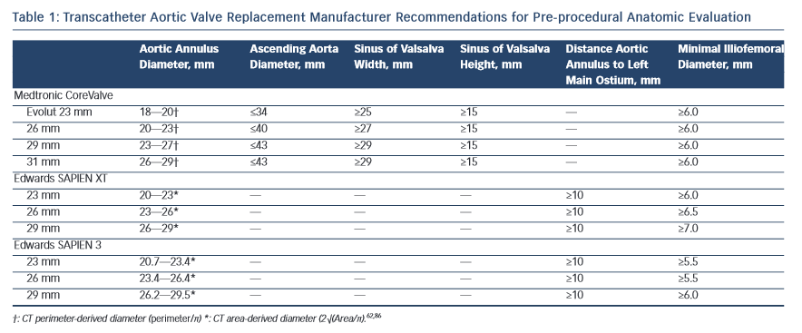
The dimensions of the aortic valve complex are critical for sizing of device (see Table 1 and Figure 1). The aortic annulus, sinuses of Valsalva, ascending aorta, coronary arteries ostia and any bypass grafts should be assessed in all patients considered for TAVR.5 While 2D echocardiography provides valuable haemodynamic information and is readily available, a MSCT-based analysis of the aortic root and vascular access sites has become a sine qua non step in the pre-operative evaluation of patients for TAVR.6 Device sizing using multi-slice computer tomography (MSCT) has been shown to offer more precise measurements of the aortic root than echocardiography and thus reduces rates of paravalvular leak (PVL),7–10 an independent predictor of post-operative mortality from 30 days to 2 years.11–20

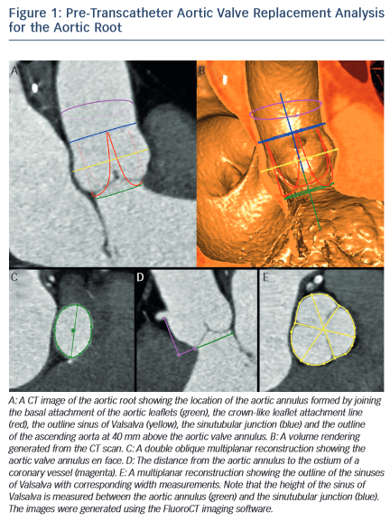
MSCT provides a precise method of evaluating aortic valve calcification (see Figure 2). Calcification is particularly important because it correlates with rates of post-intervention PVL9,21–24 It was recently demonstrated that rates of at least moderate PVL increase with left ventricular outflow tract (LVOT) calcification (OR 2.8, [95 % CI 1.2–7.0], P=0.022), and a leaflet calcium volume greater than 235 mm3 for a threshold of 850 HU (OR 2.8 [95 % CI 1.2–6.7], P=0.023).24

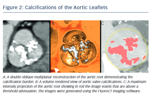
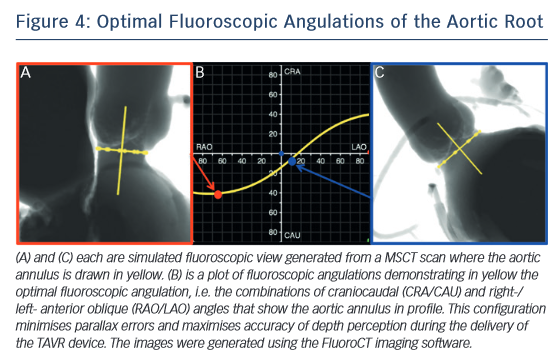
One of the most common complications of TAVR is a new left bundle branch (LBBB) or complete atrioventricular (AV) block requiring the implantation of a permanent pacemaker. The pathophysiology of these iatrogenic conduction abnormalities is the mechanical compression of the left bundle branch in the uppermost aspect of the muscular interventricular septum by the TAVR implant (see Figure 3). An increased depth of implantation is the most frequently identified predictor of LBBB with both balloon- and self-expandable prostheses.25–30 In order to obtain an optimal implantation depth, the operator must minimise parallax inherent to 2D fluoroscopy used during the implantation.31 MSCT can be used to plan optimal C-arm angulations,32–41 i.e. angulations that present the axis of the aortic root parallel to the fluoroscopic detector (see Figure 4). In a recent study,41 such optimised angulations were associated with a significant decrease in implantation time (P=0.0001), radiation exposure (P=0.02), amount of contrast (P=0.001), and risk of acute kidney injury (P=0.03).

Transcatheter Aortic Valve Replacement Manufacturer Recommendations

Article image

Image of Pre-Transcatheter Aortic Valve Replacement Analysis

Article image

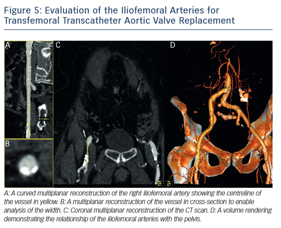
MSCT also plays a role in determining the suitability of access vessels in patients evaluated for TAVR. The majority of TAVR performed today is via the retrograde trans-femoral approach. This technique is the least invasive when compared with trans-apical, trans-aortic, trans-subclavian or trans-carotid TAVR.42-–44 While less invasive, transfemoral TAVR can result in vessel dissection, stenosis, perforation, rupture, arterio-venous fistula, pseudoaneurysm, haematoma or nerve injury.45 The main risk factors of vascular injury secondary to the insertion of the catheter sheath include the vessel diameter, calcification, and tortuosity,46,47 (see Figure 5). Contrast-enhanced MSCT provides information about these three aspects within the same imaging study. The sheath-to-vessel diameter ≥1.05–1.12 is a predictor of vascular injury when measured with CT.47,48 The use MSCT correlates with a decrease in vascular complications.46 MSCT also yields a greater predictive value for vascular complications than plain angiography.48

Figure of Calcifications of the Aortic Leaflets

Article image

Relationship of the Left Bundle Branch with the Aortic Root

Article image

Optimal Fluoroscopic Angulations of the Aortic Root

Article image

Evaluation of the lliofemoral Arteries for Transfermoral Transcatherter Aortic Valve Replacement

Article image

Image of Mitral Valve Annulus

Article image

Mitral valve

Motivated by the success of TAVR, transcatheter mitral valve procedures are seen as the next frontier of structural cardiac interventions for patients at high surgical risk. These procedures consist in the implantation of a device that repairs or replaces the native mitral valve leaflets with the goal of reducing mitral valve regurgitation. The therapies fall into four categories49; edge-to-edge repair (MitraClip), annuloplasty rings (Carillon, Mitralign, Accucinch, Cardioband), chordal implants (NeoChord, V-Chordal) and transcatheter mitral valve replacement (CardiAQ mitral valve, Fortis mitral valve, TIARA mitral valve, Tendyne mitral valve, HighLife mitral valve). These therapies are at various stages of development, from pre-clinical research to commercial availability.

Optimal Fluoroscopic Angulations of the Mitral Valve Annulus

Article image

MSCT will likely play a crucial goal in the assessment of the mitral valvular complex with these new therapies. MSCT has been investigated to assess the function and anatomy of the mitral valve. In the context of mitral regurgitation, MSCT can be used to determine the disease etiology,50,51 to quantify the severity,52,53 to describe changes in the geometry of the valvular complex54,55 and to diagnose mitral valve prolapse.56–58

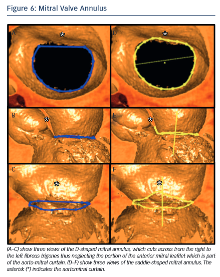
In the context of transcatheter mitral valve replacement (TMVR), MSCT has been proposed to quantify the mitral valve annulus. The mitral valve annulus is often described as either saddle-shaped or as D-shaped (see Figure 6). It has been argued that for some of the devices, in particular those that are not axially symmetrical, the D-shaped annulus may be more appropriate for sizing purposes.59,60 This question remains to be studied, but in the interim we suggest that both techniques be employed.

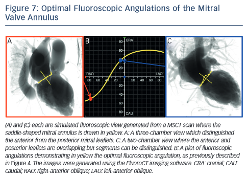
MSCT also plays a role in the optimisation of fluoroscopic viewing angles for mitral valve therapies.31,61 In particular, MSCT allows one to determine the optimal projection curve of the mitral valve annulus (see Figure 7). One may also preselect a fluoroscopic viewing angle corresponding to two-chamber view, which distinguishes mitral valve segments A1P1 from A2P2 and from A3P3 but overlaps the anterior and posterior leaflets such that A1 overlaps P1, A2 overlaps P2, and A3 overlaps P3. Conversely, one may preselect a fluoroscopic viewing angle corresponding to a three-chamber view that distinguishes the anterior from the posterior leaflets but overlaps segments A1,2,3 and separately P1,2,3.

Figure of Mitral Valve Leaflets

Article image

Image of Left atrial Appendage

Article image

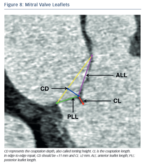
In the context of percutaneous edge-to-edge repair such as the MitraClip procedure, MSCT has been proposed for patient selection. Anatomical criteria essential for this procedure include:62,63 (1) central regurgitant jet located in the A2 and P2 mitral leaflets; (2) in functional mitral regurgitation, a coaptation length ≥2 mm and coaptation depth <11 mm (see Figure 8) and (3) in degenerative mitral regurgitation, a flail gap <10 mm and flail width <15 mm.

As new mitral valve devices undergo clinical trials, MSCT will likely play a critical role in determining patient eligibility. Further research is necessary to determine if MSCT has an impact on procedural success and clinical outcomes of these interventions.

Atrial Septal Defect and Patent Foramen Ovale

The prevalence of a patent foramen ovale (PFO) reaches 25 to 30 % of the general population. In patients having suffered a cryptogenic stroke, the incidence reaches 45.9 %,64 which suggests that PFO may play a significant role in the pathogenesis of cryptogenic stroke.65 Atrial septal defects (ASD) are the most common congenital heart defect found in adult patients.

The indications for defect closure include right ventricular volume overload with a pulmonary-to-systemic flow ratio >2:1, paradoxical embolisation and platypnea-orthodeoxia syndrome. The transcatheter treatment of PFOs and ASDs was first performed in 1975.66 Subsequent studies demonstrated that percutaneous closure has a similar rate of success when compared to surgical closure but offered reduced rates of complications and duration of hospital stay.67–69

Contrast-enhanced MSCT using a saline-chaser can be used to detect and differentiate inter-atrial shunts.70,71 and can also be used to assess disease severity.72 Ostium secundum ASDs, which represent 70 % of cases, are the only type amenable to percutaneous closure. In this context, MSCT can be useful to establish the diagnosis of sinusvenosus type ASD with partial anomalous pulmonary venous return, a condition better treated surgically.73,74

MSCT is also comparable to but less invasive than trans-esophageal echocardiography in the assessment of ASD prior to percutaneous septal occluder implantation.75

Thromboembolic Prophylaxis by Left Atrial Appendage Closure

Atrial fibrillation (AF) is the most common arrhythmia in the general population76 and is an important factor in the pathophysiology of atrial thrombus formation. Chronic anticoagulation is generally indicated in patients with AF and deemed at risk for stroke. A significant number of patients for whom thromboprophylaxis would be indicated are not eligible due to a high bleeding risk. Interestingly, studies suggest that 90 % of thrombus causing strokes in patients with AF originate from the left atrial appendage (LAA).77 This is thought to result from the presence of pectinate muscles within the LAA thus creating an appropriate milieu for blood stasis and thrombus formation. This prompted the development of surgical LAA ligation and eventually, of minimally invasive procedures.

Percutaneous transcatheter LAA closure was first described in 2002 and has since been studied in clinical trials. The Watchman Left Atrial Appendage System for Embolic Protection in Patients With Atrial Fibrillation trial (PROTECT AF trial) demonstrated that the procedure is non-inferior to warfarin therapy but resulted in higher rates of complications such as pericardial effusion.78 Four devices are currently available or under investigation: the WATCHMAN device, the Amplatzer Cardiac Plug, the WaveCrest device and the Lariat epicardial suturesnare delivery device.

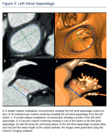
MSCT allows the anatomy of the LAA to be evaluated, which is valuable for device selection,79 assessment of procedural success and longer-term outcomes.80 It has been suggested that the perimeter-derived diameter of the LAA ostium is the most appropriate for device sizing.79 Other important parameters include the minimum and maximum diameters, as well as the waist length of the LAA ostium81 (see Figure 9). MSCT can also be used to determine optimal fluoroscopic angulation for the deployment of the LAA closure device.31

The information provided by MSCT regarding outcomes is complementary to that obtained from transoesophageal echocardiography (TEE). MSCT has a higher rate of detection than TEE for device leaks, which are defined as flow of blood within the LAA past the device.82 The localisation of leaks is also easier with MSCT than with TEE.82 Contrast-enhanced MSCT also enables the evaluation of device thrombus,83 embolisation and pericardial effusion.84,85 Further studies are necessary to provide evidence regarding the impact of MSCT imaging on outcomes of LAA closure.

Conclusions

Herein, we described the utility of MSCT in the context of structural heart interventions. With the fast development of these therapies, MSCT will likely play a role in the development of future sizing algorithms. In the future, interventional cardiologists will likely need to become imaging experts to offer their patients the most optimal outcomes from structural heart interventions.

References

  1. Turina J, Hess O, Sepulcri F and Krayenbuehl HP. Spontaneous course of aortic valve disease. Eur Heart J 1987;8:471–83.
    PubMed
  2. Nishimura RA, Otto CM, Bonow RO, et al. and Members AATF. 2014 AHA/ACC Guideline for the Management of Patients With Valvular Heart Disease: a report of the American College of Cardiology/American Heart Association Task Force on Practice Guidelines. Circulation 2014;129:e521–643.
    Crossref | PubMed
  3. Adams DH, Popma JJ, Reardon MJ, et al. and Investigators USCC. Transcatheter aortic-valve replacement with a selfexpanding prosthesis. N Engl J Med 2014;370:1790–8.
    Crossref | PubMed
  4. Mack MJ, Leon MB, Smith CR, et al. and investigators Pt. 5-year outcomes of transcatheter aortic valve replacement or surgical aortic valve replacement for high surgical risk patients with aortic stenosis (PARTNER 1): a randomised controlled trial. Lancet 2015;385:2477–84.
    Crossref | PubMed
  5. Piazza N, de Jaegere P, Schultz C, et al. Anatomy of the aortic valvar complex and its implications for transcatheter implantation of the aortic valve. Circ Cardiovasc Interv 2008;1:74–81.
    Crossref | PubMed
  6. Achenbach S, Delgado V, Hausleiter J, et al. SCCT expert consensus document on computed tomography imaging before transcatheter aortic valve implantation (TAVI)/ transcatheter aortic valve replacement (TAVR). J Cardiovasc Comput Tomogr 2012;6:366–80.
    Crossref | PubMed
  7. Willson AB, Webb JG, Labounty TM, et al. 3-dimensional aortic annular assessment by multidetector computed tomography predicts moderate or severe paravalvular regurgitation after transcatheter aortic valve replacement: a multicenter retrospective analysis. J Am Coll Cardiol 2012;59:1287–94.
    Crossref | PubMed
  8. Jilaihawi H, Kashif M, Fontana G, et al. Cross-sectional computed tomographic assessment improves accuracy of aortic annular sizing for transcatheter aortic valve replacement and reduces the incidence of paravalvular aortic regurgitation. J Am Coll Cardiol 2012;59:1275–86.
    Crossref | PubMed
  9. Schultz C, Rossi A, van Mieghem N, et al. Aortic annulus dimensions and leaflet calcification from contrast MSCT predict the need for balloon post-dilatation after TAVI with the Medtronic CoreValve prosthesis. EuroIntervention: EuroPCR in collaboration with the Working Group on Interventional Cardiology of the European Society of Cardiology 2011;7:564–72.
    Crossref | PubMed
  10. Jabbour A, Ismail TF, Moat N, et al. Multimodality imaging in transcatheter aortic valve implantation and post-procedural aortic regurgitation: comparison among cardiovascular magnetic resonance, cardiac computed tomography, and echocardiography. J Am Coll Cardiol 2011;58:2165–73.
    Crossref | PubMed
  11. Abdel-Wahab M, Zahn R, Horack M, et al. and German transcatheter aortic valve interventions registry i. Aortic regurgitation after transcatheter aortic valve implantation: incidence and early outcome. Results from the German transcatheter aortic valve interventions registry. Heart 2011;97:899–906.
    Crossref | PubMed
  12. Gotzmann M, Pljakic A, Bojara W, et al. Transcatheter aortic valve implantation in patients with severe symptomatic aortic valve stenosis-predictors of mortality and poor treatment response. Am Heart J 2011;162:238–45 e1.
    Crossref | PubMed
  13. Hayashida K, Morice MC, Chevalier B, et al. Sex-related differences in clinical presentation and outcome of transcatheter aortic valve implantation for severe aortic stenosis. J Am Coll Cardiol 2012;59:566–71.
    Crossref | PubMed
  14. Kodali SK, Williams MR, Smith CR, et al. and Investigators PT. Two-year outcomes after transcatheter or surgical aorticvalve replacement. N Engl J Med 2012;366:1686–95.
    Crossref | PubMed
  15. Moat NE, Ludman P, de Belder MA, et al. Long-term outcomes after transcatheter aortic valve implantation in high-risk patients with severe aortic stenosis: the U.K. TAVI (United Kingdom Transcatheter Aortic Valve Implantation) Registry. J Am Coll Cardiol 2011;58:2130–8.
    Crossref | PubMed
  16. Sinning JM, Hammerstingl C, Vasa-Nicotera M, et al. Aortic regurgitation index defines severity of peri-prosthetic regurgitation and predicts outcome in patients after transcatheter aortic valve implantation. J Am Coll Cardiol 2012;59:1134–41.
    Crossref | PubMed
  17. Sinning JM, Scheer AC, Adenauer V, et al. Systemic inflammatory response syndrome predicts increased mortality in patients after transcatheter aortic valve implantation. Eur Heart J 2012;33:1459–68.
    Crossref | PubMed
  18. Takagi K, Latib A, Al-Lamee R, M, Chieffo A, et al. Predictors of moderate-to-severe paravalvular aortic regurgitation immediately after CoreValve implantation and the impact of postdilatation. Catheterization and cardiovascular interventions: official journal of the Society for Cardiac Angiography & Interventions. 2011;78:432–43.
    Crossref | PubMed
  19. Tamburino C, Capodanno D, Ramondo A, et al. Incidence and predictors of early and late mortality after transcatheter aortic valve implantation in 663 patients with severe aortic stenosis. Circulation 2011;123:299–308.
    Crossref | PubMed
  20. Unbehaun A, Pasic M, Dreysse S, et al. Transapical aortic valve implantation: incidence and predictors of paravalvular leakage and transvalvular regurgitation in a series of 358 patients. J Am Coll Cardiol 2012;59:211–21.
    Crossref | PubMed
  21. John D, Buellesfeld L, Yuecel S, Mueller R, Latsios G, Beucher H, Gerckens U and Grube E. Correlation of Device landing zone calcification and acute procedural success in patients undergoing transcatheter aortic valve implantations with the self-expanding CoreValve prosthesis. JACC Cardiovasc Interv 2010;3:233–43.
    Crossref | PubMed
  22. Colli A, Gallo M, Bernabeu E, et al. Aortic valve calcium scoring is a predictor of paravalvular aortic regurgitation after transcatheter aortic valve implantation. Ann Cardiothorac Surg 2012;1:156–9.
    Crossref | PubMed
  23. Haensig M, Lehmkuhl L, Rastan AJ, et al. Aortic valve calcium scoring is a predictor of significant paravalvular aortic insufficiency in transapical-aortic valve implantation. European journal of cardio-thoracic surgery: official journal of the European Association for Cardio-thoracic Surgery 2012;41:1234–40; discussion 1240–1.
    Crossref | PubMed
  24. Jilaihawi H, Makkar RR, Kashif M, et al. A revised methodology for aortic-valvar complex calcium quantification for transcatheter aortic valve implantation. Eur Heart J Cardiovasc Imaging 2014;15:1324–32.
    Crossref | PubMed
  25. Urena M, Mok M, Serra V, et al. Predictive factors and longterm clinical consequences of persistent left bundle branch block following transcatheter aortic valve implantation with a balloon-expandable valve. J Am Coll Cardiol 2012;60:1743–52.
    Crossref | PubMed
  26. Piazza N, Onuma Y, Jesserun E, et al. Early and persistent intraventricular conduction abnormalities and requirements for pacemaking after percutaneous replacement of the aortic valve. JACC Cardiovasc Interv 2008;1:310–6.
    Crossref | PubMed
  27. Franzoni I, Latib A, Maisano F, et al. Comparison of incidence and predictors of left bundle branch block after transcatheter aortic valve implantation using the CoreValve versus the Edwards valve. J Am Coll Cardiol 2013;112:554–9.
    Crossref | PubMed
  28. Colombo A and Latib A. Left bundle branch block after transcatheter aortic valve implantation: inconsequential or a clinically important endpoint? J Am Coll Cardiol 2012;60:1753-5.
    Crossref | PubMed
  29. Calvi V, Conti S, Pruiti GP, et al. Incidence rate and predictors of permanent pacemaker implantation after transcatheter aortic valve implantation with self-expanding CoreValve prosthesis. Journal of interventional cardiac electrophysiology: an international journal of arrhythmias and pacing. 2012;34:189–95.
    Crossref | PubMed
  30. Aktug O, Dohmen G, Brehmer K, et al. Incidence and predictors of left bundle branch block after transcatheter aortic valve implantation. Int J Cardiol. 2012;160:26–30.
    Crossref | PubMed
  31. Theriault-Lauzier P, Andalib A, Martucci G, et al. Fluoroscopic anatomy of left-sided heart structures for transcatheter interventions: insight from multislice computed tomography. JACC Cardiovasc Interv 2014;7:947–57.
    Crossref | PubMed
  32. Tzikas A, Schultz C, Van Mieghem NM, et al. Optimal projection estimation for transcatheter aortic valve implantation based on contrast-aortography: validation of a Prototype Software. Catheterization and cardiovascular interventions: official journal of the Society for Cardiac Angiography & Interventions 2010;76:602–7.
    Crossref | PubMed
  33. Arnold M, Achenbach S, Pfeiffer I, et al. A method to determine suitable fluoroscopic projections for transcatheter aortic valve implantation by computed tomography. J Cardiovasc Comput Tomogr 2012;6:422–8.
    Crossref | PubMed
  34. Binder RK, Leipsic J, Wood D, et al. Prediction of optimal deployment projection for transcatheter aortic valve replacement: angiographic 3-dimensional reconstruction of the aortic root versus multidetector computed tomography. Circ Cardiovasc Interv 2012;5:247-52.
    Crossref | PubMed
  35. Cockburn J, Trivedi U, de Belder A, Hildick-Smith D. Optimal projection for transcatheter aortic valve implantation determined from the reference projection angles. Catheter Cardiovasc Interv 2012;80:973–7.
    Crossref | PubMed
  36. Dvir D, Kornowski R. Percutaneous aortic valve implantation using novel imaging guidance. Catheter Cardiovasc Interv 2010;76:450–4.
    Crossref | PubMed
  37. Gurvitch R, Wood DA, Leipsic J, et al. Multislice computed tomography for prediction of optimal angiographic deployment projections during transcatheter aortic valve implantation. JACC Cardiovasc Interv 2010;3:1157-65.
    Crossref | PubMed
  38. Kempfert J, Falk V, Schuler G, et al. Dyna-CT during minimally invasive off-pump transapical aortic valve implantation. Ann Thorac Surg 2009;88:2041.
    Crossref | PubMed
  39. Kurra V, Kapadia SR, Tuzcu EM, et al. Pre-procedural imaging of aortic root orientation and dimensions: comparison between X-ray angiographic planar imaging and 3-dimensional multidetector row computed tomography. JACC Cardiovasc Interv 2010;3:105–13.
    Crossref | PubMed
  40. Poon KK, Crowhurst J, James C, et al. Impact of optimising fluoroscopic implant angles on paravalvular regurgitation in transcatheter aortic valve replacements -- utility of three-dimensional rotational angiography. EuroIntervention 2012;8:538–45.
    Crossref | PubMed
  41. Samim M, Stella PR, Agostoni P, et al. Automated 3D analysis of pre-procedural MDCT to predict annulus plane angulation and C-arm positioning: benefit on procedural outcome in patients referred for TAVR. JACC Cardiovasc Imaging 2013;6:238–48.
    Crossref | PubMed
  42. Thourani VH, Gunter RL, Neravetla S, et al. Use of transaortic, transapical, and transcarotid transcatheter aortic valve replacement in inoperable patients. Ann Thorac Surg 2013;96:1349–57.
    Crossref | PubMed
  43. Noble S. Transapical aortic valve implantation: a reasonable therapeutic option, but not the only alternative to transfemoral approach. J Thorac Dis 2013;5:360–1.
    Crossref | PubMed
  44. Bleiziffer S, Ruge H, Mazzitelli D, et al. Survival after transapical and transfemoral aortic valve implantation: talking about two different patient populations. J Thorac Cardiovasc Surg 2009;138:1073–80.
    Crossref | PubMed
  45. Leon MB, Piazza N, Nikolsky E, et al. Standardized endpoint definitions for Transcatheter Aortic Valve Implantation clinical trials: a consensus report from the Valve Academic Research Consortium. J Am Coll Cardiol 2011;57:253–69.
    Crossref | PubMed
  46. Toggweiler S, Gurvitch R, Leipsic J, et al. Percutaneous aortic valve replacement: vascular outcomes with a fully percutaneous procedure. J Am Coll Cardiol 2012;59:113–8.
    Crossref | PubMed
  47. Hayashida K, Lefevre T, Chevalier B, et al. Transfemoral aortic valve implantation new criteria to predict vascular complications. JACC Cardiovasc Interv 2011;4:851–8.
    Crossref | PubMed
  48. Okuyama K, Jilaihawi H, Kashif M, et al. Transfemoral access assessment for transcatheter aortic valve replacement: evidence-based application of computed tomography over invasive angiography. Circ Cardiovasc Imaging 2015;8:e001995
    Crossref | PubMed
  49. Ruiz CE, Kliger C, Perk G, et al. Transcatheter Therapies for the Treatment of Valvular and Paravalvular Regurgitation in Acquired and Congenital Valvular Heart Disease. J Am Coll Cardiol 2015;66:169–83.
    Crossref | PubMed
  50. Killeen RP, Arnous S, Martos R, et al. Chronic mitral regurgitation detected on cardiac MDCT: differentiation between functional and valvular aetiologies. Eur Radiol 2010;20:1886–95.
    Crossref | PubMed
  51. Kim K, Kaji S, An Y, et al. Mechanism of asymmetric leaflet tethering in ischemic mitral regurgitation: 3D analysis with multislice CT. JACC Cardiovasc Imaging 2012;5:230–2.
    Crossref | PubMed
  52. Alkadhi H, Wildermuth S, Bettex DA, et al. Mitral regurgitation: quantification with 16-detector row CT--initial experience. Radiol 2006;238:454–63.
    Crossref | PubMed
  53. Guo YK, Yang ZG, Ning G, et al. Sixty-four-slice multidetector computed tomography for preoperative evaluation of left ventricular function and mass in patients with mitral regurgitation: comparison with magnetic resonance imaging and echocardiography. Eur Radiol 2009;19:2107–16.
    Crossref | PubMed
  54. Delgado V, Tops LF, Schuijf JD, et al. Assessment of mitral valve anatomy and geometry with multislice computed tomography. JACC Cardiovasc Imaging 2009;2:556–65.
    Crossref | PubMed
  55. Shudo Y, Matsumiya G, Sakaguchi T, et al. Assessment of changes in mitral valve configuration with multidetector computed tomography: impact of papillary muscle imbrication and ring annuloplasty. Circulation 2010;122(Suppl 11):S29–36.
    Crossref | PubMed
  56. Feuchtner GM, Alkadhi H, Karlo C, et al. Cardiac CT angiography for the diagnosis of mitral valve prolapse: comparison with echocardiography1. Radiol 2010;254:374–83.
    Crossref | PubMed
  57. Shah RG, Novaro GM, Blandon RJ, et al. Mitral valve prolapse: evaluation with ECG-gated cardiac CT angiography. AJR Am J Roentgenol 2010;194:579–84.
    Crossref | PubMed
  58. Ghosh N, Al-Shehri H, Chan K, et al. Characterization of mitral valve prolapse with cardiac computed tomography: comparison to echocardiographic and intraoperative findings. Int J Cardiovasc Imaging 2012;28:855–63.
    Crossref | PubMed
  59. Blanke P, Dvir D, Cheung A, et al. Mitral Annular Evaluation With CT in the Context of Transcatheter Mitral Valve Replacement. JACC Cardiovasc Imaging 2015;8:612–5.
    Crossref | PubMed
  60. Blanke P, Dvir D, Cheung A, et al. A simplified D-shaped model of the mitral annulus to facilitate CT-based sizing before transcatheter mitral valve implantation. J Cardiovasc Comput Tomogr 2014;8:459–67.
    Crossref | PubMed
  61. Blanke P, Dvir D, Naoum C, et al. Prediction of fluoroscopic angulation and coronary sinus location by CT in the context of transcatheter mitral valve implantation. J Cardiovasc Comput Tomogr 2015;9:183–92.
    Crossref | PubMed
  62. Ewe SH, Klautz RJ, Schalij MJ, Delgado V. Role of computed tomography imaging for transcatheter valvular repair/ insertion. Int J Cardiovasc Imaging 2011;27:1179–93.
    Crossref | PubMed
  63. Beigel R, Wunderlich NC, Kar S, Siegel RJ. The evolution of percutaneous mitral valve repair therapy: lessons learned and implications for patient selection. J Am Coll Cardiol 2014;64:2688–700.
    Crossref | PubMed
  64. Lamy C, Giannesini C, Zuber M, et al. Clinical and imaging findings in cryptogenic stroke patients with and without patent foramen ovale: the PFO-ASA Study. Atrial Septal Aneurysm. Stroke 2002;33:706–11.
    Crossref | PubMed
  65. Cheng TO. Mechanisms of platypnea-orthodeoxia: what causes water to flow uphill? Circulation 2002;105:e47.
    PubMed
  66. King TD, Thompson SL, Steiner C, Mills NL. Secundum atrial septal defect. Nonoperative closure during cardiac catheterization. JAMA 1976;235:2506–9.
    Crossref | PubMed
  67. Warnes CA, Williams RG, Bashore TM, et al. ACC/AHA 2008 Guidelines for the Management of Adults with Congenital Heart Disease: a report of the American College of Cardiology/American Heart Association Task Force on Practice Guidelines (writing committee to develop guidelines on the management of adults with congenital heart disease). Circulation 2008;118:e714–833.
    Crossref | PubMed
  68. Silversides CK, Dore A, Poirier N, et al. Canadian Cardiovascular Society 2009 Consensus Conference on the management of adults with congenital heart disease: shunt lesions. Can J Cardiol 2010;26:e70–9.
    Crossref | PubMed
  69. Baumgartner H, Bonhoeffer P, De Groot NM, et al. Task Force on the Management of Grown-up Congenital Heart Disease of the European Society of C, Association for European Paediatric C and Guidelines ESCCfP. ESC Guidelines for the management of grown-up congenital heart disease (new version 2010). Eur Heart J 2010;31:2915–57.
    Crossref | PubMed
  70. Kosehan D, Akin K, Koktener A, et al. Interatrial shunt: diagnosis of patent foramen ovale and atrial septal defect with 64-row coronary computed tomography angiography. Jpn J Radiol 2011;29:576–82.
    Crossref | PubMed
  71. White HD, Halpern EJ, Savage MP. Imaging of adult atrial septal defects with CT angiography. JACC Cardiovasc Imaging 2013;6:1342–5.
    Crossref | PubMed
  72. Osawa K, Miyoshi T, Morimitsu Y, et al. Comprehensive assessment of morphology and severity of atrial septal defects in adults by CT. J Cardiovasc Comput Tomogr 2015;9:354–61.
    Crossref | PubMed
  73. Hoey ET, Lewis G, Yusuf S. Multidetector CT assessment of partial anomalous pulmonary venous return in association with sinus venosus type atrial septal defect. Quant Imaging Med Surg 2014;4:433–4.
    Crossref | PubMed
  74. Kafka H and Mohiaddin RH. Cardiac MRI and pulmonary MR angiography of sinus venosus defect and partial anomalous pulmonary venous connection in cause of right undiagnosed ventricular enlargement. AJR Am J Roentgenol 2009;192:259–66.
    Crossref | PubMed
  75. Ko SF, Liang CD, Yip HK, et al. Amplatzer septal occluder closure of atrial septal defect: evaluation of transthoracic echocardiography, cardiac CT, and transesophageal echocardiography. AJR Am J Roentgenol 2009;193:1522–9.
    Crossref | PubMed
  76. Go AS, Hylek EM, Phillips KA, et al. Prevalence of diagnosed atrial fibrillation in adults: national implications for rhythm management and stroke prevention: the AnTicoagulation and Risk Factors in Atrial Fibrillation (ATRIA) Study. JAMA 2001;285:2370–5.
    Crossref | PubMed
  77. Blackshear JL, Odell JA. Appendage obliteration to reduce stroke in cardiac surgical patients with atrial fibrillation. Ann Thorac Surg 1996;61:755–9.
    Crossref | PubMed
  78. Reddy VY, Doshi SK, Sievert H, et al. and Investigators PA. Percutaneous left atrial appendage closure for stroke prophylaxis in patients with atrial fibrillation: 2.3-Year Follow-up of the PROTECT AF (Watchman Left Atrial Appendage System for Embolic Protection in Patients with Atrial Fibrillation) Trial. Circulation 2013;127:720–9.
    Crossref | PubMed
  79. Wang Y, Di Biase L, Horton RP, et al. Left atrial appendage studied by computed tomography to help planning for appendage closure device placement. J Cardiovasc Electrophysiol 2010;21:973–82.
    Crossref | PubMed
  80. Ismail TF, Panikker S, Markides V, et al. CT imaging for left atrial appendage closure: a review and pictorial essay. J Cardiovasc Comput Tomogr 2015;9:89–102.
    Crossref | PubMed
  81. Vaitkus PT, Wang DD, Guerrero M, et al. Left atrial appendage closure with amplatzer septal occluder in patients with atrial fibrillation: CT-based morphologic considerations. J Invasive Cardiol 2015;27:258–62.
    PubMed
  82. Jaguszewski M, Manes C, Puippe G, et al. Cardiac CT and echocardiographic evaluation of peri-device flow after percutaneous left atrial appendage closure using the AMPLATZER cardiac plug device. Catheter Cardiovasc Interv 2015;85:306–12.
    Crossref | PubMed
  83. Romero J, Husain SA, Kelesidis I, et al. Detection of left atrial appendage thrombus by cardiac computed tomography in patients with atrial fibrillation: a meta-analysis. Circ Cardiovasc Imaging 2013;6:185–94.
    Crossref | PubMed
  84. Saw J, Fahmy P, DeJong P, et al. Cardiac CT angiography for device surveillance after endovascular left atrial appendage closure. Eur Heart J Cardiovasc Imaging 2015: epub ahead of print.
    Crossref | PubMed
  85. Romero J, Cao JJ, Garcia MJ, Taub CC. Cardiac imaging for assessment of left atrial appendage stasis and thrombosis. Nat Rev Cardiol 2014;11:470–80.
    Crossref | PubMed
  86. de Araujo Goncalves P, Campos CA, Serruys PW, Garcia- Garcia HM. Computed tomography angiography for the interventional cardiologist. Eur Heart J Cardiovasc Imaging 2014;15:842–54.
    Crossref | PubMed