Article

Routine Fractional Flow Reserve Measurement after Percutaneous Coronary Intervention: The FFR-SEARCH Study

Register or Login to View PDF Permissions
Permissions× For commercial reprint enquiries please contact Springer Healthcare: ReprintsWarehouse@springernature.com.

For permissions and non-commercial reprint enquiries, please visit Copyright.com to start a request.

For author reprints, please email rob.barclay@radcliffe-group.com.
Information image
Average (ratings)
No ratings
Your rating

Abstract

Fractional flow reserve (FFR) is the mainstay of functional haemodynamic assessment of coronary artery lesions, guiding decisions in percutaneous coronary interventions (PCI). The RXi® rapid exchange FFR system, featuring an ultrathin monorail pressure microcatheter (Navvus™) has the potential to simplify PCI procedures. Data from two studies sponsored by ACIST Medical Systems evaluating the clinical utility of the microcatheter system were presented at EuroPCR, which took place over 16–19 May 2017 in Paris. Early data from the FFRStent Evaluated at Rotterdam Cardiology Hospital (FFR-SEARCH) registry have indicated that post-PCI, almost half of patients have FFR values below 0.90 even when stent placement appears fine on angiography. This registry is noteworthy for including a high proportion of unstable patients. The Assessment of Catheter-based Interrogation and Standard Techniques for Fractional Flow Reserve measurement (ACIST-FFR) study has shown that the microcatheter system provides a modestly lower FFR value compared with the traditional pressure wire, and an independent predictor of a difference between the two is the physiological severity of the lesion as measured by the Navvus microcatheter, meaning that the clinical impact of the difference is minimal for most measurements. These findings add to the growing body of evidence in support of the microcatheter FFR system and have prompted further research into optimising procedures.

Copyright Statement:

The copyright in this work belongs to Radcliffe Medical Media. Only articles clearly marked with the CC BY-NC logo are published with the Creative Commons by Attribution Licence. The CC BY-NC option was not available for Radcliffe journals before 1 January 2019. Articles marked ‘Open Access’ but not marked ‘CC BY-NC’ are made freely accessible at the time of publication but are subject to standard copyright law regarding reproduction and distribution. Permission is required for reuse of this content.

Intracoronary measurement of myocardial FFR is a simple, reliable and reproducible index of the physiological significance of coronary stenosis. In addition, FFR assessment can be used to guide PCI, improving procedural outcomes. However the impact of FFR values after PCI on clinical outcomes remains unclear.

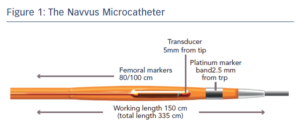
The FFR-SEARCH study aimed to determine the impact of post-PCI FFR values on long-term clinical outcomes in a prospective all-comer registry study.9 All patients who underwent successful PCI were eligible. An FFR assessment using the ACIST RXi system and Navvus microcatheter was performed after each PCI when the angiographic result was considered acceptable by the operator. Wire access to the vessel was maintained and a pressure sensor FFR microcatheter was inserted over the previously used coronary guide-wire. The Navvus microcatheter allows FFR assessment after crossing the lesion with any 0.014" guidewire of choice. A platinum marker band is located 2.5 mm from the tip and a fibre-optic sensor 2.5 mm from the marker band (5 mm from the tip).7 This gives a profile comparable to 0.022" diameter at the lesion site (see Figure 1).

The study screened 1,512 patients undergoing PCI. Of these, 512 patients were excluded: 156 because of unstable disease, 148 because of operator decision, 129 because vessels were too small and 79 for other reasons. Of the 1,000 patients who underwent assessment using the Navvus microcatheter, the wire did not cross the lesion in 28 cases, equipment failure occurred in 11 cases and two patients had an adverse response to adenosine. Of the remaining patients, 1,165 lesions had a successful post-PCI FFR measurement. Failure to cross the lesion was reported in 14 procedures, nine patients were not assessed because of unstable disease, 22 lesions were not measured because of operator decision and 28 for other reasons. In 109 lesions the vessel was too small and there was one case of equipment failure. The baseline characteristics were reported on 1000 patients and procedural results were reported on 959 patients with 1,165 lesions. The average age at baseline was 64.6 ± 11.8 years and patients were predominantly male (72.5 %). Of note, clinical presentations were evenly distributed between stable angina (30.4 %), unstable angina/non ST-elevation MI (36.7 %) and acute MI (32.9 %), i.e. >60 % of the patients involved were unstable.

Where possible, FFR was performed at least 20 mm from the most distal stent then a pullback was performed to obtain pressure gradients on the distal and proximal stent edge border as well as to assess overall drift at the ostium. The average drift value was low, 0.011 ± 0.015, and the average procedural time per lesion was 5.0 ± 1.4 minutes, which included the full duration of the adenosine infusion, showing that FFR can be used quickly to evaluate PCI results. Failure to cross the post-PCI lesion occurred in 42 of 1,207 (3.5 %) attempted lesions, mostly because of tortuosity and calcification, and there were no Navvus-related complications reported.

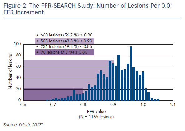
Dr Diletti presented preliminary outcome data from this study. Patientlevel analysis showed that almost half (48 %) of patients had an FFR below 0.90. Lesion level assessment showed that in 43.3 % of cases, the FFR was ≤0.90. In 7.7 % of lesions post-stenting FFR was below the threshold for ischaemia (i.e. ≤0.80). The number of lesions per 0.01 FFR increment was 660 lesions (57 %) with FFR >0.90; 505 lesions (43 %) ≤0.90; 231 lesions (20 %) ≤0.85 and 90 lesions (8 %) ≤0.80 (see Figure 2). The cut-off of 0.90 could help guide procedural optimisation.

The Navvus Microcatheter

Article image

The FFR-SEARCH Study: Number of Lesions Per 0.01 FFR Increment

Article image

The FFR-SEARCH Study: MACE at 30-day Follow-up

Article image

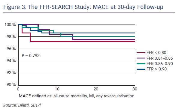
The primary outcome measure was major adverse coronary events (MACE), including all-cause mortality, Q-wave MI, target lesion revascularisation, target vessel revascularisation, stent thrombosis and any revascularisation at 30 days. Of 798 (89 %) patients with complete 30-day follow-up, MACE occurred in only 15 (1.9 %). The MACE rate for each subgroup at 30 days did not differ significantly between groups: FFR ≤0.80 2.8 %, FFR 0.81–0.85 2.6 %, FFR 0.86–0.90 2.0 % and FFR >0.90 1.5 % (see Figure 3). Although not statistically significant, it is interesting to observe the initial trend of the lower the post-PCI FFR value, the higher the reported MACE rate at 30 days. The study design intended to provide a clear message of clinical outcomes at 2 years.

Dr Diletti summarised the key points of the presentation as follows:

  • Microcatheter-based FFR post-PCI allows a safe and rapid physiological assessment of the treatment result.
  • A relevant percentage of the stented vessels have a final FFR <0.90.
  • PCI optimisation by microcatheter-based FFR may improve clinical outcome.

In the subsequent audience discussion, some experts at EuroPCR questioned whether the FFR was detecting suboptimal stent placement, or if other factors such as microcirculatory problems might be contributing. Others questioned the next step to optimising procedures. The upcoming FFR REACT randomised study aims to evaluate the clinical impact of intravascular-ultrasound-directed FFR optimisation of PCI.

References

  1. Kern MJ, Lerman A, Bech JW, et al. Physiological assessment of coronary artery disease in the cardiac catheterization laboratory: a scientific statement from the American Heart Association Committee on Diagnostic and Interventional Cardiac Catheterization, Council on Clinical Cardiology. Circulation 2006;114:1321–41.
    Crossref | PubMed
  2. Christou MA, Siontis GC, Katritsis DG, et al. Meta-analysis of fractional flow reserve versus quantitative coronary angiography and noninvasive imaging for evaluation of myocardial ischemia. Am J Cardiol 2007;99:450–6.
    Crossref | PubMed
  3. Tonino PA, De Bruyne B, Pijls NH, et al. Fractional flow reserve versus angiography for guiding percutaneous coronary intervention. N Engl J Med 2009;360:213–24.
    Crossref | PubMed
  4. van Nunen LX, Zimmermann FM, Tonino PA, et al. Fractional flow reserve versus angiography for guidance of PCI in patients with multivessel coronary artery disease (FAME): 5-year follow-up of a randomised controlled trial. Lancet 2015;386:1853–60.
    Crossref | PubMed
  5. Dattilo PB, Prasad A, Honeycutt E, et al., Contemporary patterns of fractional flow reserve and intravascular ultrasound use among patients undergoing percutaneous coronary intervention in the United States: insights from the National Cardiovascular Data Registry, J Am Coll Cardiol 2012;60:2337–9.
    Crossref | PubMed
  6. Briguori C, Nishida T, Adamian M, et al. Assessment of the functional significance of coronary lesions using a monorail catheter. J Invasive Cardiol 2001;13:279–86.
    PubMed
  7. Diletti R, Van Mieghem NM, Valgimigli M, et al. Rapid exchange ultra-thin microcatheter using fibre-optic sensing technology for measurement of intracoronary fractional flow reserve. EuroIntervention 2015;11:428–32.
    Crossref | PubMed
  8. Menon M, Jaffe W, Watson T, et al. Assessment of coronary fractional flow reserve using a monorail pressure catheter: the first-in-human ACCESS-NZ trial. EuroIntervention 2015;11:257–63.
    Crossref | PubMed
  9. Diletti R, on behalf of the Thoraxcenter Investigators: van Bommel R, Masdjedi K, van Zandvoort L, et al. Routine fractional flow reserve measurement after percutaneous coronary intervention: the FFR-SEARCH study. Presented at: EuroPCR, May 18 2017, Paris, France.
  10. NCT02577484. Assessment of Catheter-based Interrogation and Standard Techniques for Fractional Flow Reserve Measurement (ACIST-FFR), https://clinicaltrials.gov/ct2/ show/NCT02577484 (accessed 29 August 2017).
  11. Fearon WF, Chambers JW, Seto AH, et al FFR: Trials and new techniques. Primary Results of the Assessment of Catheterbased Interrogation and Standard Techniques for Fractional Flow Reserve Measurement Study: The ACIST-FFR Study. Presented at: EuroPCR, May 16-19 2017, Paris.