Article

The Value of High-definition Intravascular Ultrasound in the Setting of Complex Percutaneous Intervention

Register or Login to View PDF Permissions
Permissions× For commercial reprint enquiries please contact Springer Healthcare: ReprintsWarehouse@springernature.com.

For permissions and non-commercial reprint enquiries, please visit Copyright.com to start a request.

For author reprints, please email rob.barclay@radcliffe-group.com.
Information image
Average (ratings)
No ratings
Your rating

Abstract

A satellite symposium, sponsored by ACIST® Medical Systems, was held at EuroPCR 2016 to introduce new technologies in fractional flow reserve (FFR) and intravascular ultrasound (IVUS). FFR is the mainstay of functional haemodynamic assessment of coronary artery lesions, guiding decisions in percutaneous interventions (PCIs). The new RXi™ Rapid Exchange FFR system, featuring an ultra-thin monorail pressure microcatheter (Navvus®) has the potential to simplify PCI procedures as well as to allow the use of a workhorse coronary wire to facilitate both complex and routine FFR assessments. The system has been shown to be feasible and safe in everyday clinical practice and clinical studies. This session also introduced the first 60 MHz high definition (HDi™) IVUS system, which provides improved image quality over traditional IVUS systems, and is able to visualise bioresorbable vascular scaffold (BVS) without the need for contrast clearing.

Received:

Accepted:

Published online:

Support:The publication of this article was supported by ACIST Medical Systems.

Copyright Statement:

The copyright in this work belongs to Radcliffe Medical Media. Only articles clearly marked with the CC BY-NC logo are published with the Creative Commons by Attribution Licence. The CC BY-NC option was not available for Radcliffe journals before 1 January 2019. Articles marked ‘Open Access’ but not marked ‘CC BY-NC’ are made freely accessible at the time of publication but are subject to standard copyright law regarding reproduction and distribution. Permission is required for reuse of this content.

This presentation discussed the comparison of imaging tools from ACIST and Boston Scientific, together with optical coherence tomography (OCT). Dr Chieffo has found IVUS particularly useful in the assessment of multivessel disease and complex lesions. The importance of IVUS guidance in PCI is well established. A meta-analysis of 24,849 patients from three randomised controlled trials (RCTs) and 12 observational studies between 2005 and 2013 that compared IVUS- and angiography-guided PCI showed that IVUS was associated with a lower rate of major adverse cardiac events (odds ratio [OR] 0.79, 95 % confidence interval [CI] [0.69–0.91]; p=0.001). IVUS-guided PCI was also associated with significantly lower rates of all-cause mortality (OR 0.64; 95 % CI [0.51–0.81]; p<0.001), MI (OR 0.57; 95 % CI [0.42–0.78]; p<0.001), TVR (OR 0.81; 95 % CI [0.68–0.95]; p=0.01) and stent thrombosis (OR 0.59; 95 % CI [0.42–0.82]; p=0.002). The authors concluded that IVUS guidance is associated with a significant reduction in adverse events following PCI when compared with angiography guidance alone.4

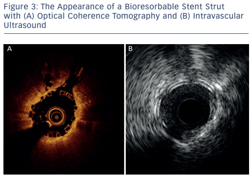
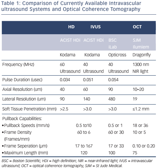
The imaging tools available in the Milan laboratory are ACIST high-definition IVUS system with a 40 and 60 MHz operation mode, Boston Scientific 40 MHz iLab™ and OCT. Table 1 shows the key features of these systems. Dr Chieffo presented three clinical cases to illustrate the differences between the systems. In the first case, highdefinition IVUS was compared to OCT in a typical patient with diffuse left anterior descending artery disease. The procedure involved the use of bioresorbable vascular scaffolds (BVS) and drug-coated balloon catheters. With these new technologies, accurate imaging tools are essential to guide procedures and to decide which stent is the most appropriate for the patient, if they are suitable for BVS and to assess the final result. Figure 3 shows the difference in images obtained by IVUS and OCT. With the40 MHz system, the strut was not easy to detect. The OCT imaging was superior in terms of determining overlapping, gaps in the BVS, branch coverage, malapposition, dissection behind the struts and asymmetric expansion with calcification. However, the use of OCT requires additional contrast and is considerably more expensive than IVUS, therefore cost-effectiveness is a factor when selecting imaging modalities.

Comparison of Currently Available Intravascular ultrasound Systems and Optical Coherence Tomography

Article image

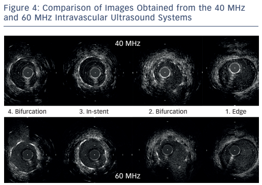
The next case was a comparison of the high-definition IVUS 60 MHz versus the Boston Scientific 40 MHz IVUS during BVS implantation. Images at different points of the affected vessel were compared. The images obtained by the 60 MHz system are not equal to the quality of OCT but are superior to the 40 MHz system and can help in decision-making. Importantly, the 60 MHz system allows imaging of the BVS without the need for contrast clearing. Another case was presented of a complex bifurcation lesion in which a drug-eluting stent was implanted. The images from the 60 MHz system are clearly superior in quality compared with the 40 MHz system (see Figure 4). In conclusion, in routine imaging, more data are needed to guide complex interventions. The 60 MHz high-definition IVUS system may become a cost-effective means of providing superior images.

The Appearance of a Bioresorbable Stent Strut with (A) Optical Coherence Tomography and (B) Intravascular Ultrasound

Article image

Comparison of Images Obtained from the 40 MHz and 60 MHz Intravascular Ultrasound Systems

Article image

References

  1. Levine GN, Bates ER, Blankenship JC, et al. 2011 ACCF/AHA/ SCAI Guideline for Percutaneous Coronary Intervention: a report of the American College of Cardiology Foundation/ American Heart Association Task Force on Practice Guidelines and the Society for Cardiovascular Angiography and Interventions. Circulation 2011;124:2574–609.
    Crossref | PubMed
  2. Task Force on Myocardial Revascularization of the European Society of Cardiology (ESC) and the European Association for Cardio-Thoracic Surgery (EACTS), European Association for Percutaneous Cardiovascular Cardiovascular Interventions (EAPCI), Wijns W, Kolh P, Danchin N, et al. Guidelines on myocardial revascularization. Eur Heart J 2010;31:2501–55.
    Crossref | PubMed
  3. Diletti R, Van Mieghem NM, Valgimigli M, et al. Rapid exchange ultra-thin microcatheter using fibre-optic sensing technology for measurement of intracoronary fractional flow reserve. EuroIntervention 2015;11:428–32.
    Crossref | PubMed
  4. Jang JS, Song YJ, Kang W, et al. Intravascular ultrasoundguided implantation of drug-eluting stents to improve outcome: a meta-analysis. JACC Cardiovasc Interv 2014;7:233–43.
    Crossref | PubMed
  5. Fearon WF, Fractional Flow Reserve-Guided Percutaneous Coronary Intervention, In: Price MJ (ed). Coronary Stenting: A Companion to Topol's Textbook of Interventional Cardiology. London: Elsevier, 2013.
  6. van Nunen LX, Zimmermann FM, Tonino PA, et al. Fractional flow reserve versus angiography for guidance of PCI in patients with multivessel coronary artery disease (FAME): 5-year follow-up of a randomised controlled trial. Lancet 2015;386:1853–60.
    Crossref | PubMed
  7. Nam CW, Mangiacapra F, Entjes R, et al. Functional SYNTAX score for risk assessment in multivessel coronary artery disease. J Am Coll Cardiol 2011;58:1211–8.
    Crossref | PubMed
  8. Menon M, Jaffe W, Watson T, Webster M. Assessment of coronary fractional flow reserve using a monorail pressure catheter: the first-in-human ACCESS-NZ trial. EuroIntervention 2015;11:257–63.
    Crossref | PubMed
  9. NCT02577484, Assessment of Catheter-based Interrogation and Standard Techniques for Fractional Flow Reserve Measurement (ACIST-FFR). Available at: https://www.clinicaltrials.gov/ct2/show/NCT02577484 (accessed 21 May 2016).
  10. Johnson NP, Jeremias A, Zimmermann FM, et al. Continuum of vasodilator stress from rest to contrast medium to adenosine hyperemia for fractional flow reserve assessment. JACC Cardiovasc Interv 2016;9:757–67.
    Crossref | PubMed